An odontoma is the most common benign odontogenic tumor of the jaws. However, many researchers consider it a hamartoma. The hamartoma is a developmental malformation rather than a true tumor. The reason behind that is its comoposition from normal tooth tissues, enamel, dentin, cementum and pulp but arranged in an abnormal pattern. Odontomas usually develop during childhood or adolescence and are discovered accidentally on routine dental radiographs. Although they are generally harmless, they can interfere with the eruption of permanent teeth and require timely diagnosis and treatment.
Types of Odontoma
Odontomas are broadly classified into two main types:
Compound Odontoma: This type consists of multiple small tooth-like structures known as denticles. Compound odontomas are more commonly present in the front region of the upper jaw. They resemble miniature teeth and are usually easier to identify on X-rays.
Complex Odontoma: A complex odontoma is composed of a disorganized mass of enamel, dentin, cementum and pulp tissue without resembling normal teeth. These lesions more frequently appear in the back region of the lower jaw.
Clinical Features
Most odontomas are asymptomatic, meaning they do not cause pain or discomfort. They are of generally detected during routine dental examinations when investigating delayed eruption of permanent teeth or retained primary teeth.
Common clinical features include:
- Delayed eruption or failure of eruption of permanent teeth.
- Retained primary teeth beyond the expected age.
- Missing teeth in the dental arch.
- Painless swelling of the jaw in larger lesions.
- Mild expansion of the surrounding bone.
- Occasionally, displacement of adjacent teeth.
- Rarely, pain or infection if the lesion becomes secondarily infected.
Radiographically, a mature odontoma appears as a well-defined radiopaque mass surrounded by a thin radiolucent (dark) rim representing the connective tissue capsule. Compound odontomas show multiple tooth-like structures, whereas complex odontomas appear as a dense irregular calcified mass.
Causes of Odontoma
The exact cause of odontoma formation remains unknown. However, several factors including following may cause it:
- Genetic predisposition.
- Trauma to primary teeth during childhood.
- Local infections affecting tooth development.
- Developmental disturbances of odontogenic tissues.
- Certain hereditary syndromes such as Gardner syndrome and Hermann syndrome.
Diagnosis
Diagnosis is based on clinical examination and radiographic findings. Common imaging required for accurate diagnosis include:
- Intraoral periapical radiographs.
- Panoramic radiographs.
- Cone Beam Computed Tomography (CBCT) for complex cases requiring detailed three-dimensional assessment.
Histopathological examination after surgical removal confirms the diagnosis.
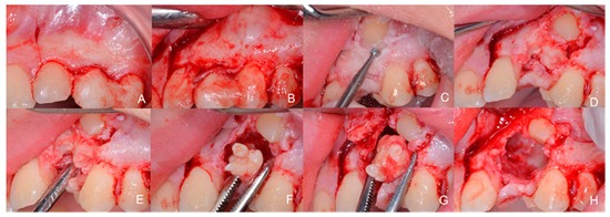
Treatment
The treatment of choice for odontoma is conservative surgical removal. The procedure is relatively simple because the lesion is usually well encapsulated and can be separated easily from the surrounding bone.
If an impacted (unerupted) permanent tooth is associated with the odontoma, the dentist or oral surgeon evaluates its eruption potential after removal. In many young patients, the impacted tooth erupts naturally. In other cases, dentists recommend orthodontic (Braces) treatment to guide the tooth into its proper position.
Postoperative healing is generally excellent and recurrence is extremely rare.
Prognosis
The prognosis of odontoma is highly favorable. Since it is a benign lesion with minimal recurrence risk, early diagnosis and treatment usually result in complete recovery. Regular dental check-ups during childhood and adolescence are important because routine radiographs can detect odontomas before they cause complications such as delayed tooth eruption, crowding, or misalignment of teeth.
References
Madani, Mohammad Ishaaq Ahmed; Gilani, Ashar Eqbal; Majumder, Dipen; Huidrom, Dinabati; Khare, Ankita. An Uncommon Case of Hamartomatous Growth (Odontome) in the Right Mandibular Region with Encroachment of the Inferior Alveolar Nerve. Current Trends in Dentistry, 2025; 2(2):117-120, DOI: 10.4103/CTD.CTD_17_25
Mazur M, Di Giorgio G, Ndokaj A, Jedliński M, Corridore D, Marasca B, Salucci A, Polimeni A, Ottolenghi L, Bossù M, et al. Characteristics, Diagnosis and Treatment of Compound Odontoma Associated with Impacted Teeth. Children. 2022; 9(10):1509. https://doi.org/10.3390/children9101509
Read our full disclaimer.